Catalog Number: CG-0156
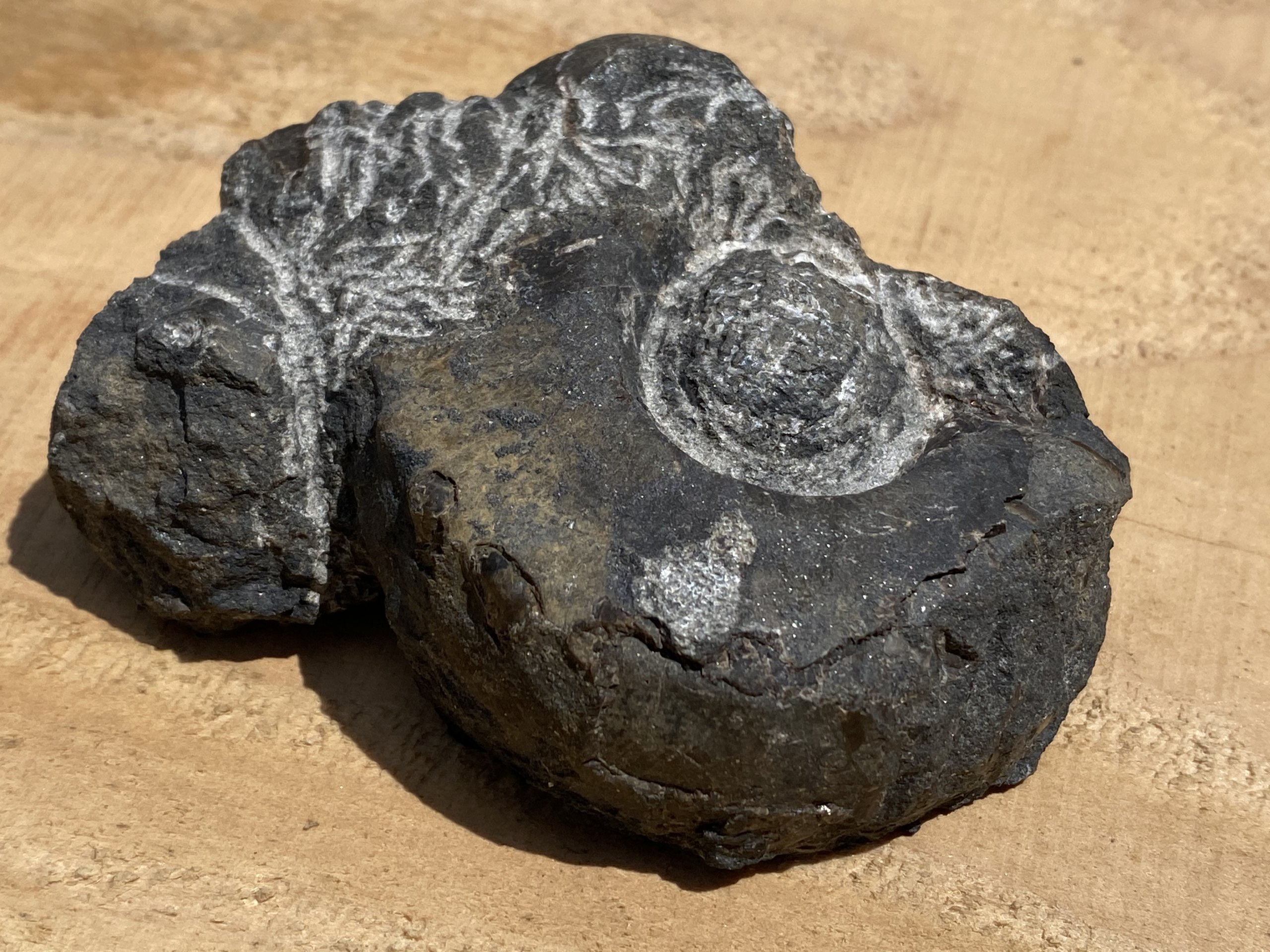
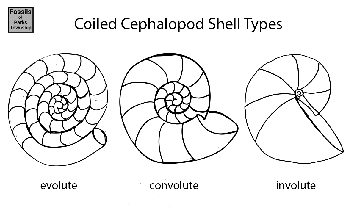
If I find a large convolute cephalopod in local Brush Creek Limestone, it’s usually the genus Metacoceras. Each find is a unique opportunity to study the genus as a whole. It’s the luck of the draw that determines how each will present itself, once a rock hammer breaks open a larger limestone boulder. In this instance, a large shell was present, showing the nodes on the shoulder. More interesting was that most of the shell material was intact. This gave me an opportunity to try to expose the rest of it.
There was a large mass of shell material from one or more secondary creatures that laid on top of the specimen. Using an air scribe, I slowly removed this material and exposed the umbilical area. The area has a surprise, a fossil brachiopod shell had rested inside of the round depression in the center of the shell whorl. As I first found it, only the top of the shell was exposed. I continued to remove the attached matrix, and it became apparent that this was an entire brachiopod shell. I took extra care not to break the shell as I worked, and this resulted in an attractive specimen.
Continued Prep and Discovered Specimen Crushing
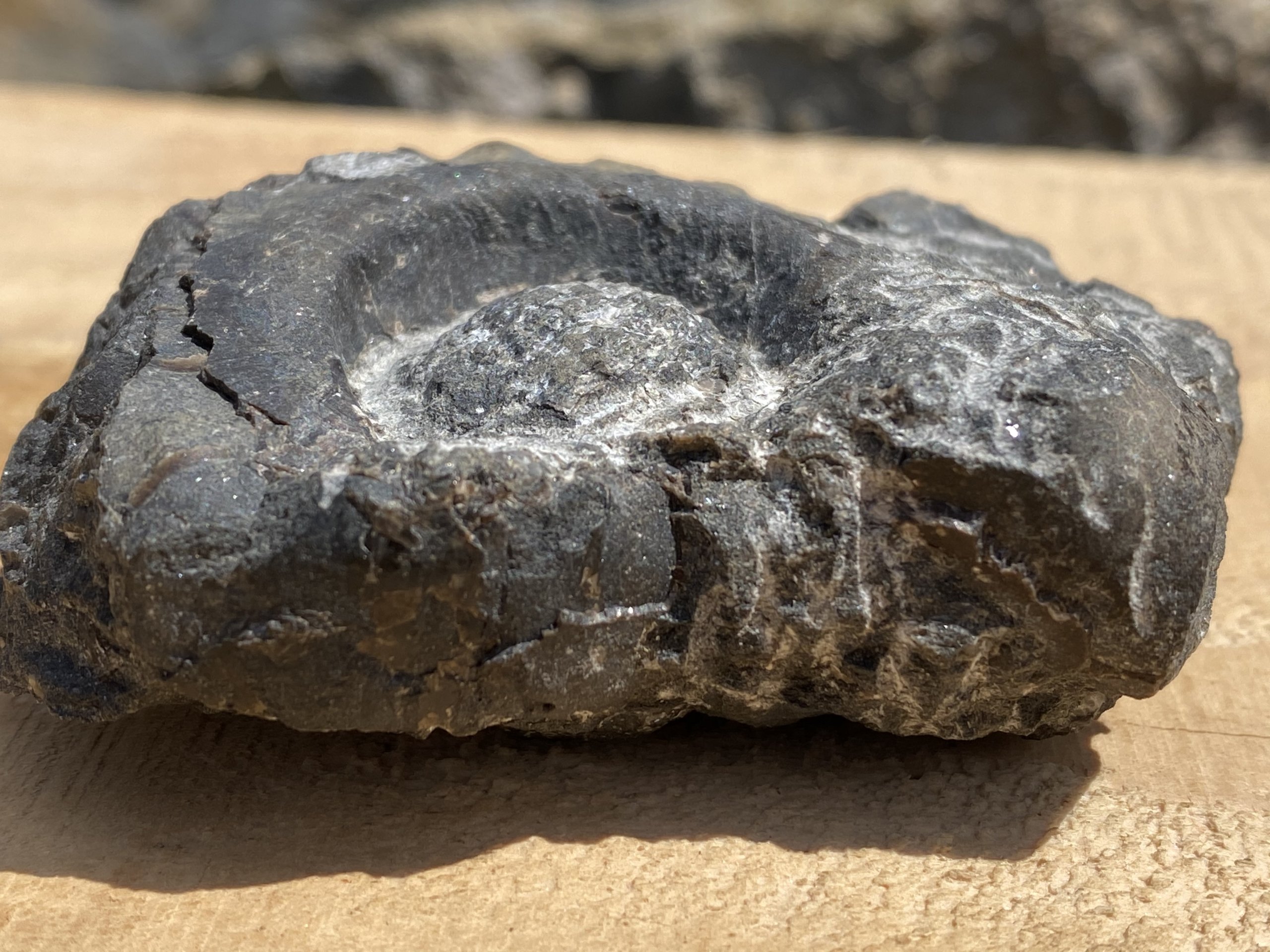
I was able to expose the umbilical wall, however, I did not make it to the umbilical seam. The brachiopod shell seemed to take up all of the available space in this area. The umbilical diameter was roughly 30mm as measured from the top of the umbilical shoulder. This tapered inwards towards the center. The exposed wall height was 8mm at its highest. The whorl flank is 16mm on average, of the large portion.
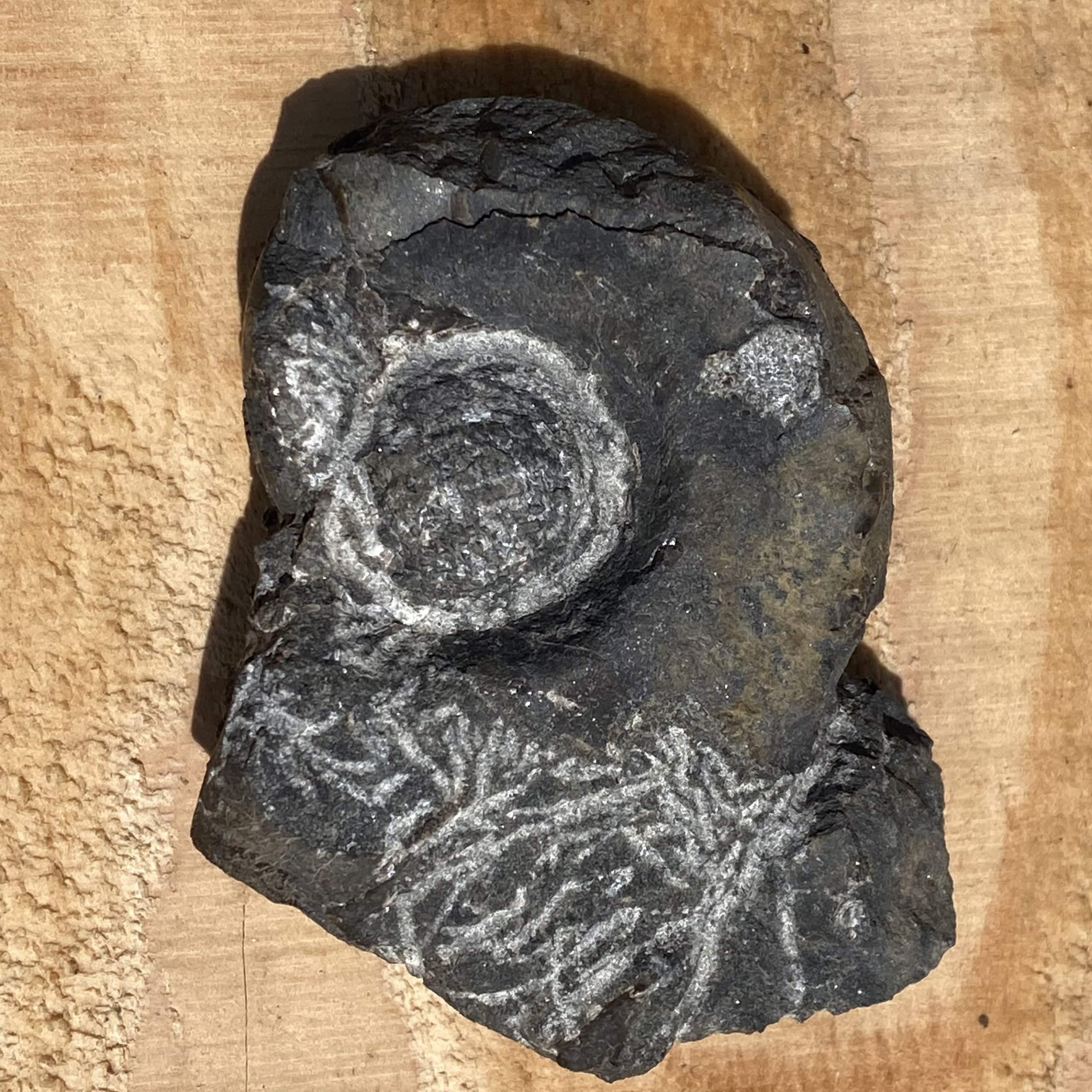
Half of the whorl has not been crushed. In profile, it is apparent where the shell caves in on itself, from venter inwards towards the umbilical area. The side opposite of the one shown in Figure 1 shows crushing, where the ventrolateral shoulder is visible as a ridge. There is more matrix to be removed in the crushed area, however, it was decided to cut the rock in this area first, then use an air scribe to finish the work.

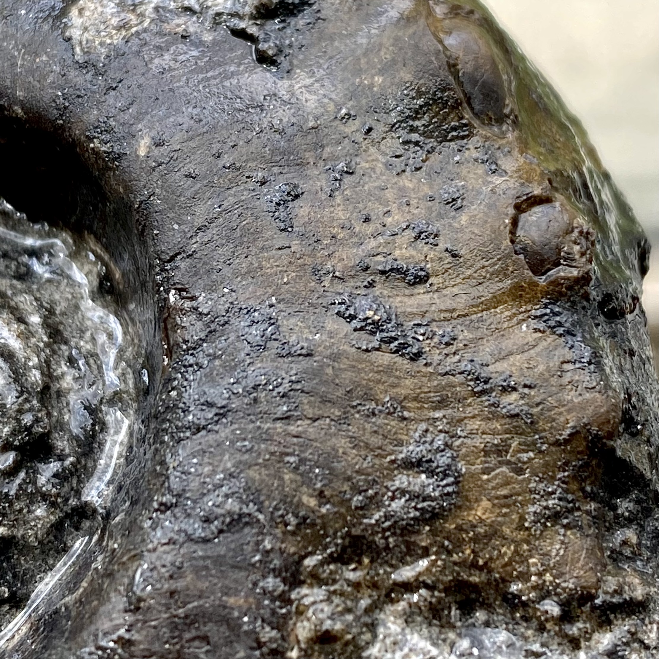
Preserved Shell Detail
One common challenge is to not destroy the shell when prepping recovering specimens. Fossil shells of Metacoceras are smooth on the inside and textured on the outside. The outside sticks more to the matrix, and attempting to remove the matrix from the outside often results in the shattering of the shell material. This shell material is mostly made of calcite. Calcite is more prone to shattering.
Once exposed, the shell preserves the natural growth pattern of Metacoceras. The pattern is a sweeping S pattern. Each line or groove is representative of the edge of the shell as the creature’s mantle secreted shell material while growing the shell. Used often in identification, this pattern helps differentiate several different types of mollusks.
More reading online about Metacoceras
- 1883, Hyatt, A., Proceedings of the Boston Society of Natural History V. 22, Genera of Fossil Cephalopods, Boston Society of Natural History. P. 268-269
- 2020, Godlesky, C., Metacoceras, Fossils of Parks Township.