Crosswise cells or rays are typical but often hidden components of the vascular tissue in plans. Fossil plants occur in the shale layers below the Brush Creek limestone. These sometimes preserve incredible detail, including detailed carbon films from plants living over 300 million years ago. While shale plant fossils are excellent, I also find plant fossils in limestone. When found, they are usually gold in color, perhaps made of the mineral pyrite or marcasite.
These are a rare find here. Having woody plant material fall into the sea, sink into the sediment, and achieve preservation is unlikely. But it does happen. The three specimens I have found to date are all gold in color.
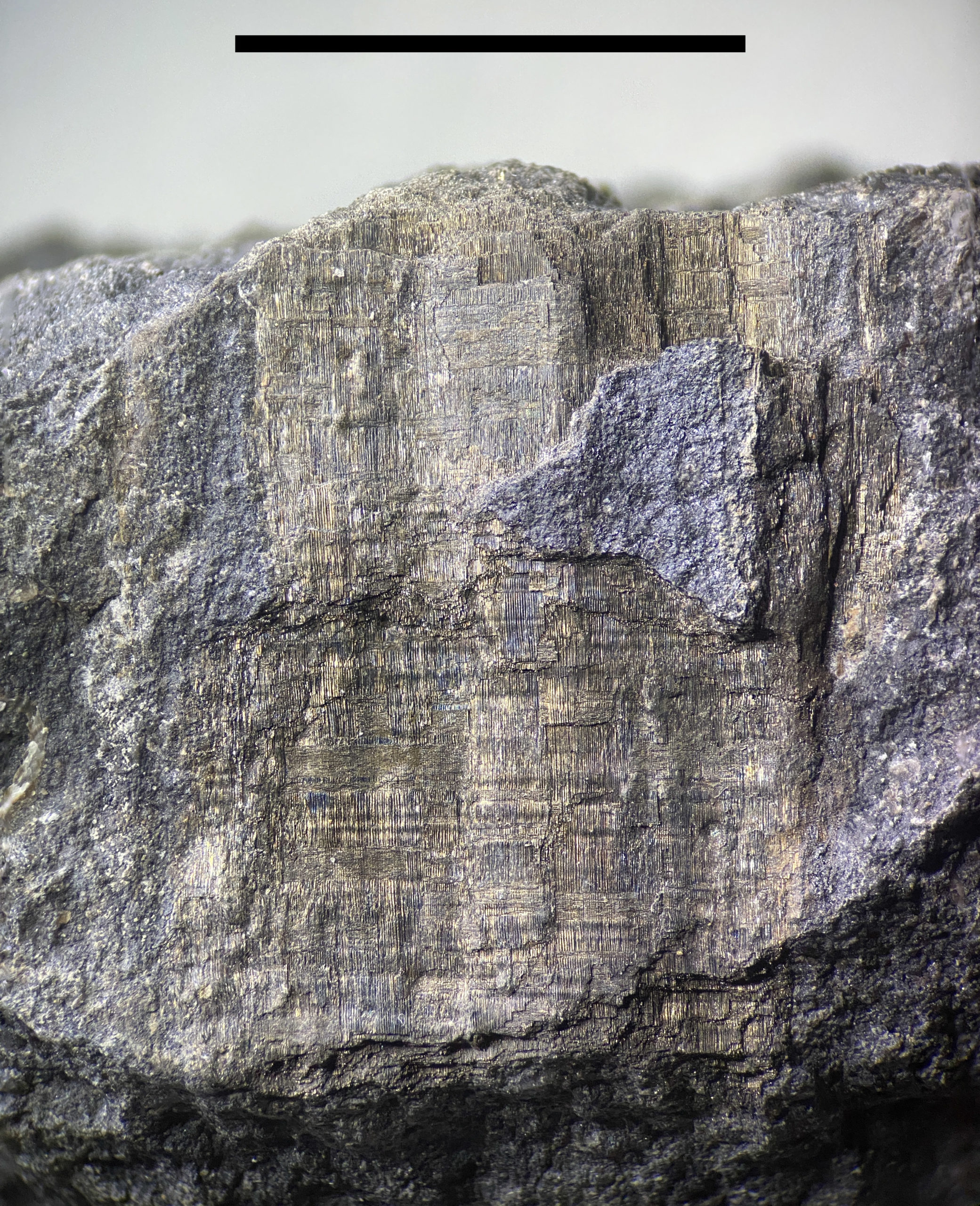
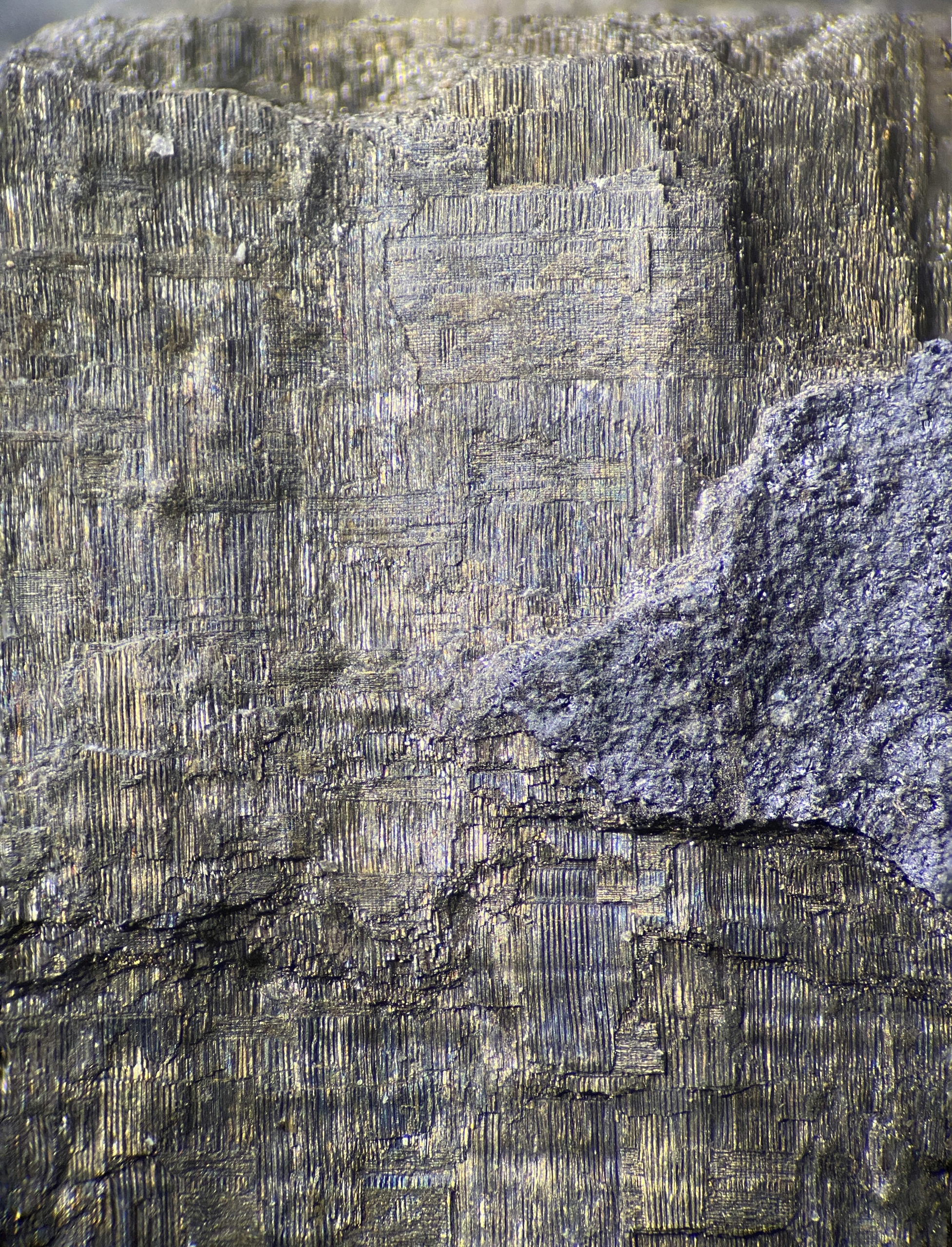
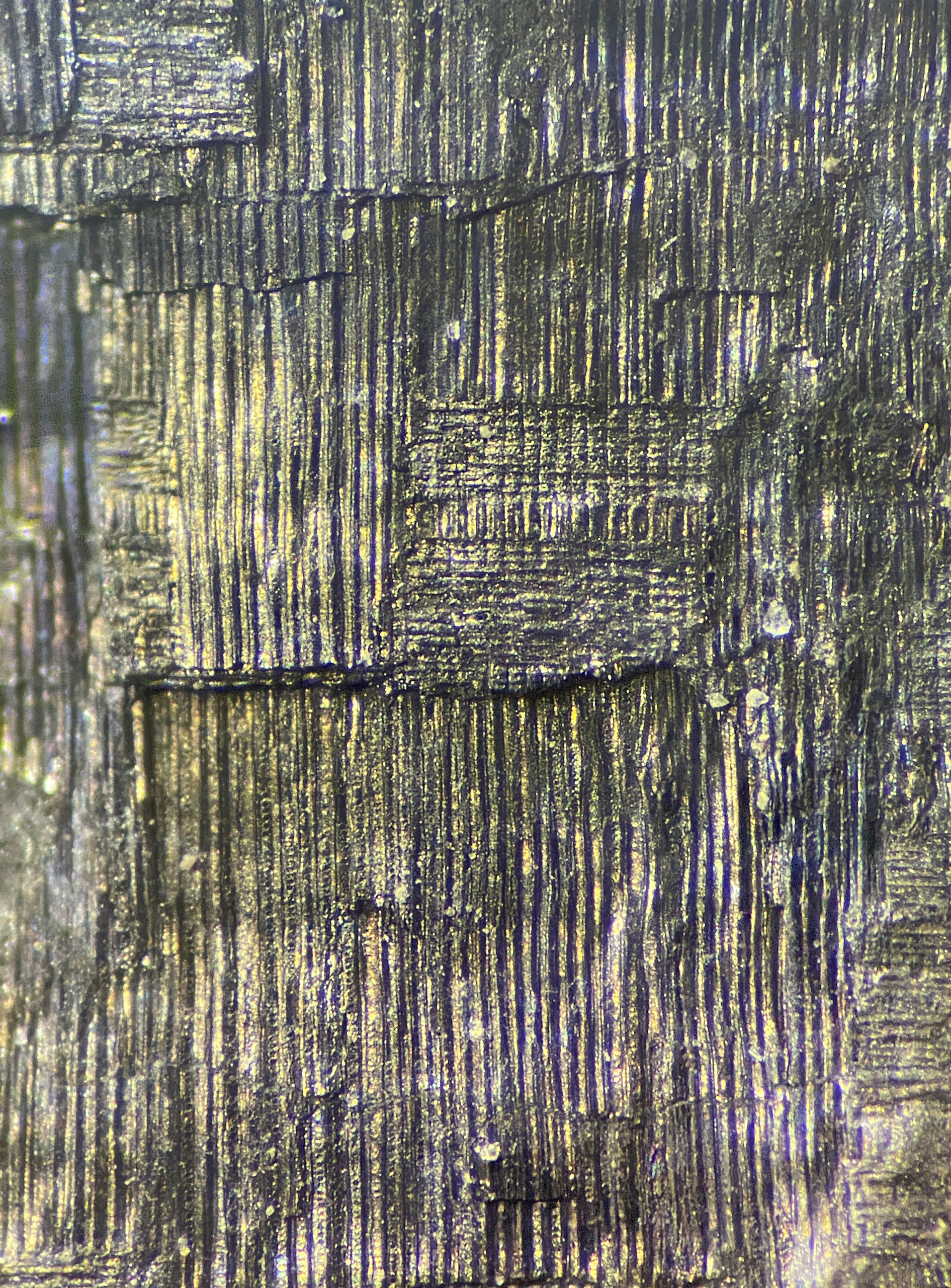
Preservation shows Crosswise cells.
Looking at the specimens under a microscope reveals preserved vascular tissue. The orientation for most is in the direction of growth, but many are in a perpendicular direction. These rays or crosswise oriented cells help support the growth structure of the other cells and help move food and water deeper into the wood. They also provide more strength and flexibility.
Microscope Photography of the Cells
I photographed the following specimens at several different depths and combined them into one focus-stacked image.

References
- Britannica.com – The anatomy and organization of wood
- Chan J., 2012, Microtubule and cellulose microfibril orientation during plant cell and organ growth. J Microsc. 247(1):23-32.
- Chafe, S.C., Wardrop, A.B. (1972) Fine structural observations on the epidermis. I. The epidermal cell wall. Planta 187, 269-278