Catalog Number: CG-0038
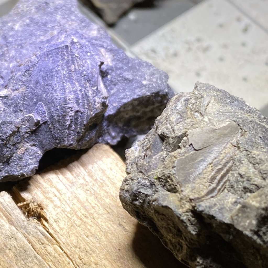
While sorting through the endless piles of fossil pieces I have set near the lab microscope, I found another piece of a Petalodus Tooth. This is the most incomplete of the teeth I’ve found to date, with only a microscopic tooth chip left behind. However, this helps with microscopic views of the interior layers of the tooth, as seen below. M.C. Hansen says in his 1985 paper, Systematic Relationships of Petalodontiform Chondrichthyans, that two things characterize teeth within the order of Petalodontiformes. These are external morphology and microscopic anatomy (Hansen 1985). Before modern methods of slicing and viewing fossil Petalodont teeth were possible, finding specimens like this one offered a clear view into the inner anatomy of the tooth. Beyond the enamel so to say.
The images below are poor from a microscopic view. I hope to perform some focus stacking and produce better images in the future.
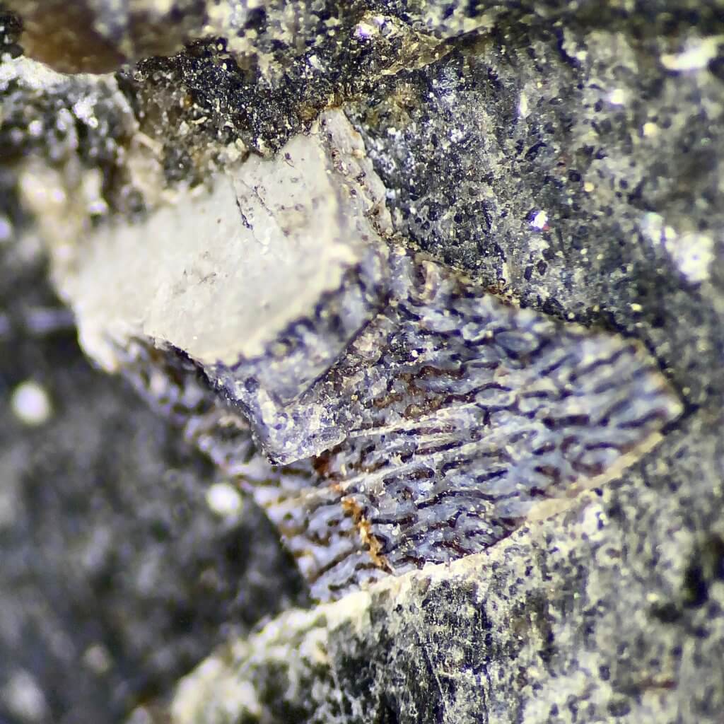
I may have identified this as a tooth fragment in the past and set it aside, however, the memory of that happening escapes me. I am mostly sure however that this was brought in during 2019. Another best-defined feature in this specimen is the impression of the distal crown tongue, which has been buried in limestone matrix in previous examples I have found. This specimen has 5 distinct ridges visible.
References
- Hansen, M.C., 1985, Systematic Relationships of Petalodontiform Chondrichthyans